For decades, scientists have been studying intriguing “gravity holes,” which are enormous depressions in the Earth’s crust where the effects of gravity are significantly lower than average.
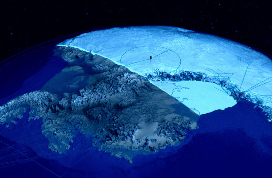
It’s an especially pertinent phenomenon in the Antarctic, a region that has seen significant changes not just due to global warming, but far longer-term climate changes spanning tens of millions of years — long before the emergence of humans and their environmentally disastrous footprint on the planet. The effects of gravity are particularly weak beneath the icy continent when accounting for our planet’s rotation, the result of slow rock movements deep beneath the ice.
As detailed in a new paper published in the journal Scientific Reports, University of Florida geophysics professor Alessandro Forte and Paris Institute of Earth Physics researcher Petar Glišović found that these rock movements are correlated to major changes in Antarctica’s climate, suggesting how the area’s gravity shifts may have allowed its ice sheets to grow.
The pair created a detailed map of the Antarctic’s “gravity hole” to study how it changed over millions of years, using a wealth of global earthquake recordings from across the planet.
“Imagine doing a CT scan of the whole Earth, but we don’t have X-rays like we do in a medical office,” said Forte in a statement. “We have earthquakes. Earthquake waves provide the ‘light’ that illuminates the interior of the planet.”
Using computer models, the team reconstructed the state of Antarctic’s gravity hole 70 million years ago, around the time when dinosaurs still roamed the Earth. They determined that the hole has gained strength over tens of millions of years, coinciding with major changes in the continent’s climate system and the widespread formation of glaciers, which in turn, had sweeping effects on sea levels the acidity of our planet’s oceans.
While the findings aren’t a definitive causal link between the two — rock movements and shifting gravity causing ice to grow — Forte and Glišović are hoping to test whether sea level changes may be directly influenced by this strengthening gravity hole.
“How does our climate connect to what’s going on inside our planet?” Forte asked rhetorically in the statement. “If we can better understand how Earth’s interior shapes gravity and sea levels, we gain insight into factors that may matter for the growth and stability of large ice sheets.”
More on gravity holes: Scientists Intrigued by “Gravity Hole” at Bottom of Ocean
The post Antarctica’s Gravity Hole Growing Stronger, Scientists Find appeared first on Futurism.