Researchers have uncovered new insights into how the brain controls eating behavior, which could have significant implications for tackling obesity. Their study, published in Nature Metabolism, shows that certain neurons in the brains of mice can either promote eating due to hunger or suppress eating for pleasure, depending on the circumstances. These findings offer a deeper understanding of how different types of eating are regulated in the brain and could lead to new treatments for obesity.
The motivation behind this study stems from the need to understand the dual nature of eating: eating out of necessity (because of hunger) and eating for pleasure (often linked to the consumption of calorie-dense, sugary, or fatty foods). While hunger-driven eating is essential for survival, pleasure-driven eating is often associated with overeating and the development of obesity, along with related metabolic disorders.
Despite the significant public health implications of obesity, the neural mechanisms that control these different types of eating behaviors have remained largely unclear. This study aimed to shed light on these mechanisms, specifically focusing on a group of neurons identified by the proenkephalin marker in the diagonal band of Broca, a region of the mouse brain.
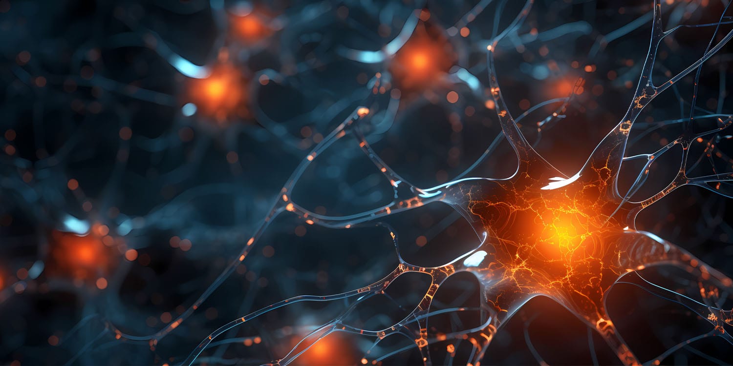
The research team focused on neurons in the diagonal band of Broca (DBB) of male mice. These neurons are marked by a protein called proenkephalin, which is involved in the body’s opioid system and has been linked to feeding behavior in previous studies. The researchers wanted to determine whether these neurons play a role in regulating hunger-driven and pleasure-driven eating differently.
To do this, they used a variety of techniques, including optogenetics, a method that allows scientists to control neurons with light. By activating or deactivating specific groups of neurons, they could observe how these changes affected the eating behavior of the mice. The researchers also used advanced imaging techniques to track the activity of these neurons in real-time as the mice were exposed to different types of food under different conditions, such as during hunger or when presented with high-fat, high-sugar (HFHS) foods.
In their experiments, they found that DBB proenkephalin neurons could be divided into two subgroups based on where their projections led within the brain. One group of neurons projected to the paraventricular nucleus of the hypothalamus (PVH), a brain region involved in hunger regulation. The other group projected to the lateral hypothalamus (LH), which is known to be involved in pleasure-driven eating.
When the neurons projecting to the PVH were activated, the mice were more likely to eat regular chow, especially when they were hungry. This suggests that these neurons help promote hunger-driven feeding, making sure that the body gets the energy it needs when it is running low on fuel.
On the other hand, the neurons projecting to the LH had the opposite effect when it came to pleasure-driven eating. Activation of these neurons decreased the consumption of high-fat, high-sugar foods, even when these foods were freely available and typically very appealing to the mice. This indicates that these neurons can suppress pleasure-driven eating, potentially acting as a brake to prevent overeating.
“Ideal feeding habits would balance eating for necessity and for pleasure, minimizing the latter,” said co-corresponding author Yong Xu, a professor of pediatrics and associate director for basic sciences at the USDA/ARS Children’s Nutrition Research Center at Baylor College of Medicine. “In this study we identified a group of neurons that regulates balanced feeding in the brain.”
Further investigation showed that these effects were linked to how the neurons were wired into different brain circuits. The PVH-projecting neurons were specifically active when food was presented during periods of fasting, which aligns with the idea that they promote hunger-driven eating. Conversely, the LH-projecting neurons became active when the mice were presented with high-calorie foods, but instead of encouraging eating, they inhibited it. This was a surprising discovery because it was previously thought that neurons in this area of the brain would promote pleasure-driven feeding rather than suppress it.
“A subset of DBB-Penk neurons that projects to the paraventricular nucleus of the hypothalamus is preferentially activated upon food presentation during fasting periods, facilitating hunger-driven feeding,” Xu said. “On the other hand, a separate subset of DBB-Penk neurons that projects to a different brain region, the lateral hypothalamus, is preferentially activated when detecting high-fat, high-sugar (HFHS) foods and inhibits their consumption. This is the first study to show a neural circuit that is activated by a reward, HFHS, but leads to terminating instead of continuing the pleasurable activity.”
When the researchers disabled the entire population of DBB proenkephalin neurons in some mice, the results were dramatic. These mice, when given the choice between regular chow and high-fat, high-sugar foods, ate less chow and more of the HFHS diet, leading to rapid weight gain and the onset of obesity-related metabolic issues. This finding underscores the importance of these neurons in maintaining a balance between hunger-driven and pleasure-driven eating.
The study concludes that these DBB proenkephalin neurons play a critical role in controlling eating behavior by either promoting hunger-driven eating or suppressing pleasure-driven eating. The balance between these two types of eating is crucial for maintaining a healthy body weight and metabolism. The researchers suggest that disruptions in this balance could contribute to obesity and other metabolic disorders.
“Our findings indicate that the development of obesity is associated with impaired function of some of these brain circuits in mice,” Xu said. “We are interested in further investigating molecular markers within the circuits that could be suitable targets for treatment of human diseases such as obesity.”
The study, “Distinct basal forebrain-originated neural circuits promote homoeostatic feeding and suppress hedonic feeding in male mice,” was authored by Hailan Liu, Jonathan C. Bean, Yongxiang Li, Meng Yu, Olivia Z. Ginnard, Kristine M. Conde, Mengjie Wang, Xing Fang, Hesong Liu, Longlong Tu, Na Yin, Junying Han, Yongjie Yang, Qingchun Tong, Benjamin R. Arenkiel, Chunmei Wang, Yang He, and Yong Xu.


