A new study suggests that damage to the brainstem, the part of the brain that controls essential functions like breathing and heart rate, may be responsible for the long-lasting physical and mental health issues seen in patients who experienced severe COVID-19. Using powerful, ultra-high-resolution MRI scanners, researchers from the Universities of Cambridge and Oxford observed abnormalities in the brainstems of individuals who had been hospitalized with severe COVID-19 early in the pandemic.
These abnormalities are believed to result from inflammation, and they appear to correlate with symptoms such as breathlessness, fatigue, and anxiety. The findings, published in the journal Brain, could help explain why some people experience prolonged symptoms after recovering from COVID-19.
The researchers conducted this study to better understand the long-term effects of severe COVID-19 on the brain. When the pandemic first began, many patients reported lingering symptoms long after they had recovered from the initial infection, a phenomenon that has come to be known as long COVID. These symptoms often include fatigue, breathlessness, and anxiety, but the underlying cause of these issues remained unclear. Early post-mortem studies showed evidence of inflammation in the brainstems of patients who had died from COVID-19, leading scientists to suspect that the brainstem might play a role in long COVID.
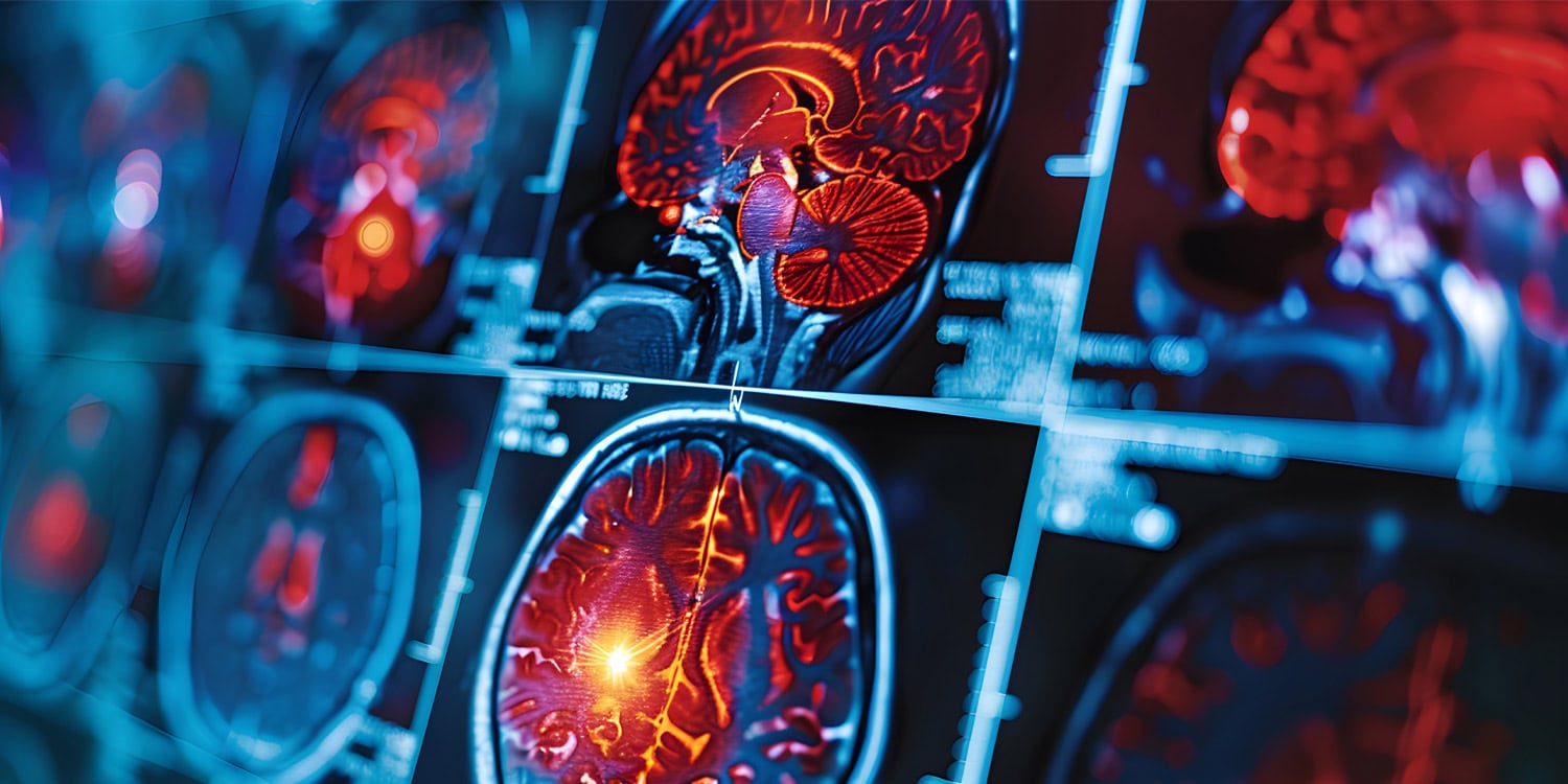
However, it had previously been impossible to study this inflammation in living people, because of the brainstem’s small size and location deep within the brain. With the advent of 7-Tesla (7T) MRI scanners, which can image the brain in much finer detail than conventional scanners, the researchers hoped to identify any lasting damage to the brainstem in survivors of severe COVID-19.
“Things happening in and around the brainstem are vital for quality of life, but it had been impossible to scan the inflammation of the brainstem nuclei in living people, because of their tiny size and difficult position,” said first author Catarina Rua, from Cambridge’s Department of Clinical Neurosciences. “Usually, scientists only get a good look at the brainstem during post-mortem examinations. But with 7T scanners, we can now measure these details. The active immune cells interfere with the ultra-high magnetic field, so that we’re able to detect how they are behaving. Cambridge was special because we were able to scan even the sickest and infectious patients, early in the pandemic.”
“The brainstem is the critical junction box between our conscious selves and what is happening in our bodies,” said Professor James Rowe, also from the Department of Clinical Neurosciences, who co-led the research. “The ability to see and understand how the brainstem changes in response to COVID-19 will help explain and treat the long-term effects more effectively.”
The study involved 30 participants who had been hospitalized with severe COVID-19 during the early stages of the pandemic, before vaccines were available. These participants were scanned using powerful 7-Tesla MRI scanners several months after they had recovered and been discharged from the hospital. For comparison, the researchers also scanned a group of 51 healthy individuals who had no history of COVID-19 infection. The participants were recruited from two sites: the Wolfson Brain Imaging Centre at Cambridge and the Wellcome Centre for Integrative Neuroimaging at Oxford.
The researchers used a specific type of MRI technique called quantitative susceptibility mapping (QSM), which can measure tiny changes in brain tissue by detecting the magnetic properties of certain materials like iron. QSM is particularly useful for detecting inflammation and other changes in brain structure. In this study, the researchers focused on four key regions of the brainstem: the midbrain, pons, medulla, and superior cerebellar peduncle. They were particularly interested in finding out whether these regions showed any abnormalities in the COVID-19 patients, as these areas are involved in functions like breathing and anxiety regulation.
The research team also collected clinical data from the COVID-19 patients, including measures of disease severity during their hospital stay, such as C-reactive protein (CRP) levels (a marker of inflammation) and the duration of their hospital admission. They also assessed the patients’ mental health and physical recovery using questionnaires that measured symptoms of anxiety and depression, as well as overall functional ability.
The MRI scans revealed that the COVID-19 survivors had significant abnormalities in their brainstems, particularly in the medulla oblongata and the pons, which are critical regions for regulating breathing, heart rate, and other basic bodily functions. The researchers found that these abnormalities were consistent with a neuroinflammatory response, suggesting that the patients’ immune systems had overreacted to the virus and caused damage to the brainstem. Importantly, these changes were still present several months after the patients had recovered from the acute phase of the infection.
One of the most striking findings was that the level of inflammation in the brainstem was correlated with the severity of the patients’ COVID-19 infection and their recovery outcomes. Patients who had experienced more severe cases of COVID-19, as measured by longer hospital stays and higher levels of CRP, showed greater brainstem abnormalities. Moreover, the brainstem regions that were most affected are known to be involved in regulating the sensation of breathlessness, which could explain why many of the patients continued to feel short of breath long after their infection had cleared.
“The fact that we see abnormalities in the parts of the brain associated with breathing strongly suggests that long-lasting symptoms are an effect of inflammation in the brainstem following COVID-19 infection,” said Rua. “These effects are over and above the effects of age and gender, and are more pronounced in those who had had severe COVID-19.”
The study also found that the abnormalities in the brainstem were linked to mental health issues, such as anxiety and depression. This is significant because the brainstem is responsible for monitoring breathlessness and fatigue, sensations that are closely linked to anxiety. The researchers suggested that the damage to the brainstem could explain why some COVID-19 survivors experience ongoing mental health issues, even after the physical symptoms of the infection have subsided.
“Mental health is intimately connected to brain health, and patients with the most marked immune response also showed higher levels of depression and anxiety,” said Rowe. “Changes in the brainstem caused by COVID-19 infection could also lead to poor mental health outcomes, because of the tight connection between physical and mental health.”
While this study offers valuable insights into the potential brain-based mechanisms of long COVID symptoms, it has some limitations. First, the sample size was relatively small, with only 30 COVID-19 patients included in the study. This limits the ability to generalize the findings to all COVID-19 survivors, especially those who had less severe infections or were asymptomatic. The patients included in the study were also all hospitalized during the early stages of the pandemic, before vaccines were available, so it is unclear whether the findings would apply to people who contracted COVID-19 later or who had milder cases.
Another limitation is that the study only captured a single point in time—several months after the patients had recovered. Future research should track COVID-19 survivors over longer periods to see whether the brainstem abnormalities improve, worsen, or remain stable over time. Additionally, while the study focused on the brainstem, other parts of the brain may also play a role in the long-term effects of COVID-19, and these areas should be explored in future studies.
Looking ahead, the researchers hope that their findings will pave the way for further investigations into the neurological effects of COVID-19 and other viral infections. By better understanding how inflammation in the brain affects physical and mental health, scientists may be able to develop more targeted treatments for long COVID and related conditions. The use of advanced MRI techniques like 7-Tesla scanners could also help monitor the effectiveness of treatments for other brain disorders, such as multiple sclerosis and dementia, where inflammation plays a key role.
The study, “Quantitative susceptibility mapping at 7 T in COVID-19: brainstem effects and outcome associations,” was authored by Catarina Rua, Betty Raman, Christopher T. Rodgers, Virginia F. J. Newcombe, Anne Manktelow, Doris A. Chatfield, Stephen J. Sawcer, Joanne G. Outtrim, Victoria C. Lupson, Emmanuel A. Stamatakis, Guy B. Williams, William T. Clarke, Lin Qiu, Martyn Ezra, Rory McDonald, Stuart Clare, Mark Cassar, Stefan Neubauer, Karen D. Ersche, Edward T. Bullmore, David K. Menon, Kyle Pattinson, and James B. Rowe on behalf of the Cambridge NeuroCOVID group, the CITIID-NIHR COVID-19 BioResource Collaboration and the Oxford CMORE-NEURO group.