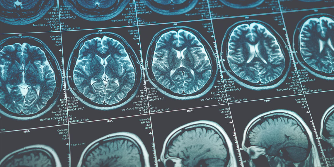
A recent neuroimaging study conducted in China suggests that a higher body mass index is associated with reduced brain volume and increased damage to white matter. Published in Health Data Science, the study found that individuals younger than 45 with a high body mass index had brain volumes resembling those of people 12 years older, highlighting the potential impact of obesity on brain health.
Obesity is defined by excessive body fat accumulation, which raises the risk of numerous health issues, including heart disease, diabetes, and certain cancers. It has become a widespread public health concern, with the World Health Organization reporting that over one billion people worldwide are classified as obese. Obesity’s prevalence continues to climb in both developed and developing nations, driven by factors like higher consumption of energy-dense foods and increasingly sedentary lifestyles.
Body mass index, a commonly used measure, helps define obesity by dividing a person’s weight in kilograms by their height squared (kg/m²). A body mass index of 30 or higher indicates obesity, while 25 to 29.9 is classified as overweight. Given the growing rates of obesity, researchers, including study lead author Han Lv, sought to investigate its potential long-term effects on brain health.
Study author Han Lv and his colleagues hypothesized that high body mass index (i.e., obesity) might have adverse effects on brain health in the long run. They conducted a neuroimaging study to observe the associations between structural and functional characteristics of the brain and body mass index.
To test this idea, they conducted a neuroimaging study as part of the Kailuan Study, a long-term cohort study of adults in Kailuan, Hebei Province, China. Begun in 2006, this research tracks a wide range of health markers over time, including body mass index and neuroimaging data, providing a rich basis for exploring potential body mass index effects on the brain.
The researchers focused on data from 1,074 participants who had completed at least one magnetic resonance imaging scan since 2020. These participants, ranging in age from 25 to 83, had body mass index data gathered across a 16-year period, allowing researchers to examine cumulative body mass index exposure. On average, each participant had five clinical assessments during the study period, which included measurements of body mass index and brain imaging. The average participant age was 55, and 44% of the sample were women.
The study analyzed participants’ brain structures in relation to their cumulative body mass index. Researchers divided participants into three groups based on body mass index: high, medium, and low. They found that participants in the high body mass index group tended to have smaller brain volumes and reduced cerebrospinal fluid volume, with differences in brain volume averaging around 8-9 milliliters when compared to those with lower body mass index values.
In younger adults, the brain volume differences were even more pronounced. Among participants under 45, those with high body mass index had brain volumes around 14-18 milliliters smaller than those with low body mass index values. The volume of cerebrospinal fluid in high body mass index participants was also reduced by about 18 milliliters. The observed changes roughly correspond to an estimated 12 years of accelerated brain aging among the high body mass index group compared to the low body mass index group.
Additionally, the researchers performed a genetic analysis called Mendelian randomization, which helps establish causal relationships by using genetic data as a basis for examining long-term body mass index’s effects on brain structure.
The Mendelian randomization analysis supported a genetic causal relationship between high body mass index and reduced gray matter volume. The genetic analysis linked high body mass index to specific brain changes, suggesting that body mass index can directly influence brain structure, not just through lifestyle factors but also through inherent biological links. Notably, high body mass index was associated with increased density in the brain’s projection fibers, which are involved in long-distance communication within the brain and control motor and sensory functions.
“High BMI is associated with a smaller brain volume, larger volume of WM lesions (damage to white matter of the brain), and abnormal microstructural integrity in projection fibers [disruptions or alterations in the structural quality of long-range nerve fibers that connect different regions of the brain]. For adults younger than 45 years, BMI should be maintained below 26.2 kg/m2 for better brain health,” the study authors concluded.
The study sheds light on the link between body mass index and structural characteristics of the brain. However, the neuroimaging data were taken only once, leaving room for further research to assess long-term changes over multiple time points.
Additionally, the study relied on body mass index as an indicator of body fat, though body mass index alone may not always reflect body composition accurately. Finally, since the genetic analysis was conducted on participants of European ancestry, the results might not fully generalize to the entire population in the study. Future studies should consider longitudinal imaging and body composition measures to address these limitations.
The paper, “Association between Body Mass Index and Brain Health in Adults: A 16-Year Population-Based Cohort and Mendelian Randomization Study,” was authored by Han Lv, Na Zeng, Mengyi Li, Jing Sun, Ning Wu, Mingze Xu, Qian Chen, Xinyu Zhao, Shuohua Chen, Wenjuan Liu, Xiaoshuai Li, Pengfei Zhao, Max Wintermark, Ying Hui, Jing Li, Shouling Wu, and Zhenchang Wang.


