A recent study published in The Journal of Neuroscience sheds light on how obesity can disrupt reproductive health in males by altering brain circuitry. The research used mice fed a high-fat diet to mimic human obesity and found that it caused chronic changes in brain connections. These changes led to reduced testosterone, lower sperm count, and diminished libido in the mice. The study provides evidence that obesity weakens communication between the brain circuits that control both feeding and reproduction, potentially explaining the link between obesity and reproductive issues in men.
The researchers were motivated to conduct this study because while it is well-known that obesity lowers testosterone in men, which impacts various functions like muscle mass, cognition, and reproductive health, the exact mechanisms by which obesity causes these changes are not fully understood. This knowledge gap is significant because obesity-related reproductive issues are becoming increasingly common.
Obese men often suffer from low testosterone levels, reduced sperm count, and poor sperm quality. This study aimed to understand how chronic obesity alters brain circuitry to produce these effects, hoping that understanding the mechanisms might eventually lead to treatments or interventions that can address reproductive dysfunction in obese men.
“A long-term goal of my research is to identify the molecular and cellular mechanisms that regulate reproductive function, which is necessary for the survival of the species,” said corresponding author Djurdjica Coss, a professor of biomedical sciences and associate vice chancellor for research at the University of California, Riverside School of Medicine.
“My research is significant to individuals struggling with unexplained infertility. Currently, 1 in 8 couples experience infertility and require assisted reproductive technologies to have a child. It is also important for the survival of endangered species, whose preservation depends on reproductive assistance, and for our food supply, as agricultural animals increasingly suffer from infertility due to modern farming practices. The studies in my lab may help identify new treatments and strategies to alleviate conditions that contribute to the rising infertility rates in both humans and animals.”
“An increase in infertility in the Western world has coincided with the growing prevalence of obesity, which now affects 35% of individuals in the United States,” Coss explained. “Obese individuals have a higher incidence of various diseases, including reproductive disorders. Obesity is known to lower testosterone in men, impacting muscle mass and cognition, as well as reproductive function by reducing sperm numbers and decreasing libido. The global rise in obesity may partly explain the decline in sperm counts that has been reported in the media. However, the mechanistic links between obesity and infertility remain unclear.”
To mimic the effects of human obesity, the researchers used male mice fed a high-fat diet. These mice were compared to a control group that was fed a standard diet. After 12 weeks, the researchers measured the levels of luteinizing hormone (LH), a hormone critical for testosterone production and sperm development, to assess the impact of the high-fat diet on reproductive function.
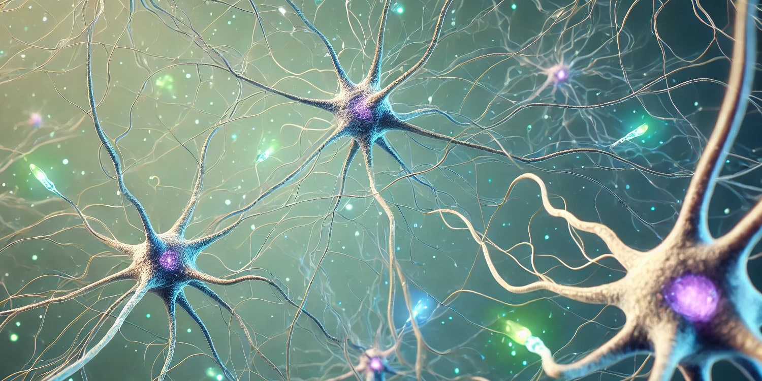
The team specifically focused on two groups of neurons in the hypothalamus: proopiomelanocortin (POMC) neurons, which play a role in regulating energy balance and food intake, and kisspeptin neurons, which are crucial for controlling the release of gonadotropin-releasing hormone (GnRH) and, consequently, LH.
The researchers found that obesity caused significant changes in the brain’s reproductive circuitry. In obese mice, LH pulse frequency was reduced, leading to lower testosterone levels and reduced sperm counts. While the reproductive system retained its ability to function normally under direct stimulation, the chronic effects of obesity suppressed the activity of kisspeptin neurons, which are essential for triggering the release of GnRH and LH.
The team observed a reduction in the number of receptors on kisspeptin neurons that respond to αMSH, a molecule produced by POMC neurons that normally helps coordinate feeding and reproductive functions. This reduction in receptor availability weakened the communication between POMC and kisspeptin neurons, leading to a suppression of kisspeptin activity and reduced LH secretion.
Another key finding was that glutamatergic signaling, which is thought to help synchronize kisspeptin neuron activity, was reduced in obese mice. This decrease in glutamatergic input likely contributed to the impaired reproductive function observed in the high-fat diet group.
Despite these changes, the researchers found that when kisspeptin neurons were artificially activated through chemogenetic techniques, the LH response was greater in obese mice than in the control group, suggesting that the kisspeptin neurons were not permanently damaged but were being suppressed by the effects of obesity.
“The extent of changes was surprising,” Coss told PsyPost. “Our studies demonstrated that the brain is the primary site for obesity’s impact on reproductive function, specifically populations of neurons that regulate the reproductive hormone axis and food intake. Neurons in the brain are connected and communicate with each other via synapses. Neurons that regulate food intake and energy expenditure interact with neurons that regulate reproduction to coordinate their functions, since reproduction is an energy-demanding process.”
“Using mice fed a high-fat diet to mimic human obesity, my team found that obesity causes chronic changes in the brain. We showed that the brains of the chronically fat mice have fewer connections between neurons and a downregulation — a reduction in the number — of receptors that normally inform the brain that enough energy is available to stop food intake. This may explain why we don’t halt excessive calorie intake and why it is difficult to lose weight. We counted the number of synapses (connections) in the neurons that regulate reproduction in the brain and identified fewer synapses in the obese mice. We still don’t know exactly how this happens, but now, after identifying specific neuronal populations and specific synaptic molecules that are affected by obesity, we can design future studies to define mechanisms or identify treatments.”
Interestingly, the research team previously reported that while male mice fed a high-fat diet experienced significant reductions in LH levels, testosterone, and sperm counts, female mice did not show the same degree of reproductive impairment. This suggests that females may be more resistant to the negative reproductive effects of obesity compared to males.
“Our previous studies have shown that females gain weight more slowly, and that even after weight gain, females are somewhat protected from the significant negative effects of obesity,” Coss explained. “This also matches observations in humans, where males are more susceptible to cardiovascular disease and metabolic syndrome in obesity.”
“However, we expected more changes in females after weight gain. We now think that females are more accustomed to weight fluctuations due to pregnancy or the need to store more energy while nursing. For now, that is only a hypothesis, and we are investigating possible mechanisms that provide protection to females.”
One limitation of the study is that it was conducted in mice, and although mouse models are commonly used in scientific research, they may not fully replicate the complexity of human physiology. The researchers also point out that the timing and duration of obesity may play a role in how severely the brain’s reproductive circuits are affected. Chronic, long-term obesity may lead to more pronounced changes in brain function compared to short-term obesity.
Future studies could explore how different lengths of high-fat diet exposure impact reproductive health and whether interventions like diet changes or exercise can reverse the negative effects of obesity on the brain. Further research is needed to explore these differences and understand why females may be more resistant to the reproductive impacts of obesity.
“Our goal is to understand the etiology of disease in order to either prevent it or identify treatments,” Coss said. “Our studies analyzing sex differences may help us identify protective mechanisms in one sex and use it to protect a weaker sex (in obesity case, male).”
The study, “Obesity Alters POMC and Kisspeptin Neuron Cross Talk Leading to Reduced Luteinizing Hormone in Male Mice,” was authored by Pedro A. Villa, Rebecca E. Ruggiero-Ruff, Bradley B. Jamieson, Rebecca E. Campbell, and Djurdjica Coss.