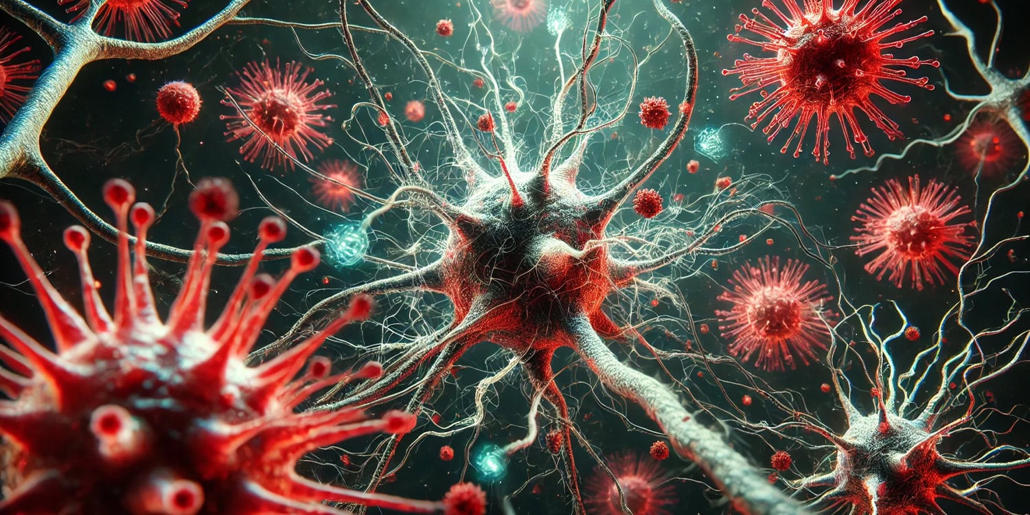
A new study published in Nature Microbiology has revealed that SARS-CoV-2, the virus responsible for COVID-19, evolves more rapidly in the brain than in the lungs, possibly explaining some of the neurological symptoms associated with the disease. This finding may provide insight into the mysterious phenomenon of long COVID and might eventually lead to targeted treatments that specifically address the virus in the central nervous system.
COVID-19 is primarily known for attacking the respiratory system, but it has also been linked to a wide range of other symptoms, including neurological effects such as loss of smell, memory issues, and what has come to be known as “brain fog.” These symptoms are part of a broader set of long-term effects that some patients experience, collectively referred to as long COVID. Despite the widespread neurological impact, the mechanisms behind how the virus affects the brain remain unclear.
SARS-CoV-2 has been shown to spread to other parts of the body, including the heart, gastrointestinal tract, and central nervous system. However, the factors that enable the virus to infect these distant tissues are not fully understood. The new study aimed to explore whether the virus mutates differently in various parts of the body, particularly in the brain and lungs, and to understand the role these mutations play in the virus’s ability to infect different tissues.
“COVID-19 has long been associated with a number of neurological symptoms, including loss of taste/smell and ‘brain fog.’ In addition, a subset of people experience post-acute sequelae of COVID-19 (PASC), otherwise known as ‘long COVID’, which also has a number of neurological symptoms,” said co-corresponding author Judd Hultquist, an assistant professor of medicine and microbiology-immunology at Northwestern University Feinberg School of Medicine.
“It is still unknown if these symptoms are caused by infection of cells in the central nervous system and brain, by immune dysfunction following infection, or by some combination of both. We started this study to understand how we might prevent infection of brain and hopefully then prevent some of these neurological symptoms.”
The study was conducted using mouse models infected with SARS-CoV-2. Mice were vaccinated with different types of vaccines, or not vaccinated at all, before being exposed to the virus. Five days after infection, the researchers collected samples from both the lungs and the brain. They used a technique called whole-genome sequencing to analyze the viral RNA in these samples, which allowed them to track how the virus evolved in different parts of the body.
In addition to examining the virus in vaccinated versus unvaccinated mice, the researchers also looked specifically at changes in the spike protein. This protein is critical because it allows the virus to bind to and enter human cells, and mutations here have been shown to affect how easily the virus spreads. The researchers paid particular attention to a region of the spike protein called the furin cleavage site, which plays a key role in the virus’s ability to infect different types of cells.
The researchers found that SARS-CoV-2 evolved differently in the brain compared to the lungs. In the lungs, the virus looked similar to the original strain used to infect the mice. However, in the brain, the virus had accumulated more mutations, particularly in the spike protein. These mutations often disrupted the furin cleavage site.
One of the key findings was that viruses with these spike protein mutations were better at infecting the brain. When the researchers used these brain-adapted viruses to directly infect the brains of other mice, the mutations persisted. Interestingly, when the brain-adapted virus spread back to the lungs, it tended to revert to its original form. This suggests that different parts of the body create different selective pressures on the virus, influencing how it evolves.
“In brief, we discovered that SARS-CoV-2 was much better at infecting cells in the brain if it mutated a specific region of its outer spike protein,” Hultquist told PsyPost. “The spike protein determines how the virus enters a cell and these mutations forced the virus to enter cells using one particular pathway. This suggests that we can prevent infection of the brain by targeting this pathway, which may help alleviate some neurological symptoms of COVID-19.”
The researchers also found that vaccination did not significantly alter how the virus evolved in the brain compared to the lungs. Whether the mice were vaccinated or not, the virus displayed greater genetic diversity in the brain. This was a surprising discovery, as the researchers initially hypothesized that vaccination would limit viral mutations across all tissues. Instead, it appears that immune-privileged areas of the body, like the brain, may allow the virus to evolve in ways that wouldn’t be possible in the lungs, where immune responses are stronger.
“The vaccination status didn’t really determine the virus evolution, but we observed differences in the virus sequence in the brain versus the lung,” said Justin Richner, assistant professor of microbiology and immunology at the University of Illinois Chicago and co-corresponding author of the paper. “That really set us on a totally unexpected trajectory.”
The mutations that accumulated in the brain also made the virus less virulent, meaning it caused less severe disease. This was a surprising discovery because it suggests that while the virus might be better suited to infecting the brain, it could become less dangerous in doing so. Despite this, the researchers were concerned that these mutations could eventually spread back to the respiratory system, where the virus could regain its ability to cause severe illness and potentially spread to others.
“Potentially, this could be a source of novel variants of concern,” Richner explained. “It could be that the virus is using these different tissue sites to evolve new mutations, and then those can traffic back into the respiratory tract and spread throughout the population.”
“This finding suggests that the vaccines are still important because the only way the virus reaches these distal tissues is if it establishes an infection and is able to replicate in the body,” he added. “The vaccines are important to prevent the virus from reaching some of those distal tissues and undergo diversification.”
Like all studies, this research had its limitations. First, it was conducted in mice, which, while useful for modeling human disease, do not perfectly mimic human infections. The researchers acknowledged that the way the virus mutates and behaves in human tissues might differ. Additionally, they only studied a few days’ worth of viral evolution in mice. While this was enough to detect significant changes in the virus, a longer study might reveal more about how these mutations affect disease progression over time.
“It is important to point out that all of our studies were conducted in mice,” Hultquist noted. “It is yet to be seen if SARS-CoV-2 has similar requirements for infecting cells in the human brain. If these requirements are the same, we are likely several years off from seeing this work translated into the clinic.”
Another limitation was the focus on just two organs—the lungs and the brain. While these are critical sites for understanding COVID-19, the virus can also affect other tissues, such as the heart and kidneys. Future studies could examine how SARS-CoV-2 evolves in these other tissues and whether similar patterns of mutation occur.
The study opens up several new avenues for research. One major question is how the virus travels from the lungs to the brain and back again. Understanding the pathways the virus uses to move between tissues could reveal new targets for therapies. Additionally, the researchers hope to explore whether the mutations found in the brain are linked to the neurological symptoms observed in COVID-19 patients, such as brain fog and memory loss. If so, this could lead to more targeted treatments for these symptoms.
“There are some small molecule inhibitors of the pathways that the SARS-CoV-2 uses to enter the brain,” Hultquist explained. “Our future work will be to determine if they are effective at preventing infection of the brain in mice and to see if they alter the presentation or outcome of disease.”
“This research was only made possible through a cross-institutional collaboration with the lab of Dr. Justin Richner at the University of Illinois-Chicago,” Hultquist added. “Breaking down barriers and bringing people together is what stimulates new ideas and research breakthroughs. We are really thankful for all of the efforts to unite scientists in response to the COVID-19 pandemic, which had a measurable impact on patient care and saved lives.”
The study, “Evolution of SARS-CoV-2 in the murine central nervous system drives viral diversification,” was authored by Jacob Class, Lacy M. Simons, Ramon Lorenzo-Redondo, Jazmin Galván Achi, Laura Cooper, Tanushree Dangi, Pablo Penaloza-MacMaster, Egon A. Ozer, Sarah E. Lutz, Lijun Rong, Judd F. Hultquist, and Justin M. Richner.


