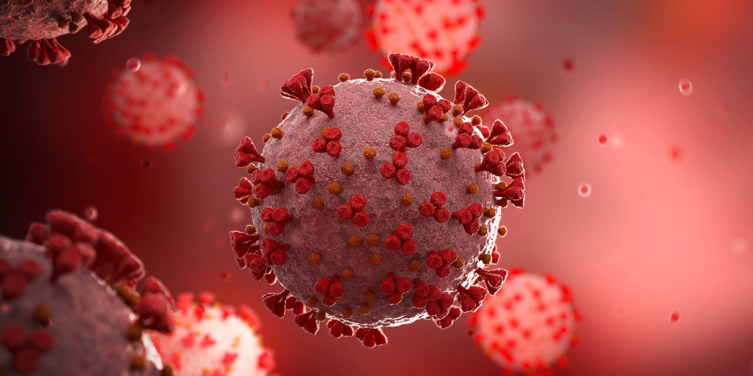
A new study published in Scientific Reports sheds light on long-term neurological consequences of COVID-19. Researchers found that individuals who had anosmia (the loss of smell) during COVID-19 showed alterations in brain functionality and even physical structure during recovery. This study is among the first to link COVID-19-related loss of smell to significant brain changes.
COVID-19, caused by the SARS-CoV-2 virus, has been primarily known for its impact on the respiratory system. However, over time, many patients, even those with mild cases, reported cognitive issues such as memory problems, confusion, and difficulties with concentration, which raised concerns about the virus’s effects on the brain. Neurological symptoms like headaches, brain fog, and loss of smell emerged as common issues for COVID-19 survivors.
Anosmia, the loss of smell, became one of the earliest and most recognizable symptoms of COVID-19, often occurring suddenly. While most patients recovered their sense of smell after a few weeks, some experienced longer-lasting olfactory dysfunctions. Previous research also suggested that loss of smell could signal broader neurological involvement in diseases like Alzheimer’s and Parkinson’s. Given the commonality of anosmia in COVID-19 and its potential implications for brain health, the researchers sought to explore whether loss of smell during COVID-19 was associated with any measurable brain changes in recovering patients.
“Our laboratory studies the neurobiological mechanisms underlying complex social behavior and decision-making. During the pandemic, it was very challenging to halt our experimental activities due to health restrictions,” said study author Pablo Billeke of the Center for Research in Social Complexity at the University for Development in Chile.
“In this context and given the early reports of neurological symptoms in patients affected by COVID-19, we wanted to contribute from our unique perspective to understanding the potential damage caused by SARS-CoV-2 infection in the central nervous system. This led us to initiate this study, in which we evaluated recovered COVID-19 patients using structural and functional magnetic resonance imaging while they performed decision-making and cognitive control tasks, as well as tracking their evolution with electroencephalography.”
To investigate these possible brain changes, the research team recruited 100 adults in Santiago, Chile, who had recovered from respiratory infections between February 2020 and May 2023. The final sample included 73 participants who had confirmed cases of COVID-19 (the remaining participants had respiratory infections caused by other agents, as confirmed by multiple negative PCR tests). The team used a combination of tests and brain scans across two sessions to assess these participants’ brain function and structure.
The participants ranged in age from 19 to 65, and none had severe cases of COVID-19 that required ventilators or intensive care. The study specifically excluded anyone with neuropsychiatric disorders or severe brain injuries, ensuring that the observed effects could be linked to their infection rather than prior conditions.
In behavioral tests, participants with a history of anosmia displayed more impulsive decision-making compared to those who did not lose their sense of smell. These individuals tended to change their choices more rapidly after receiving negative feedback, particularly in tasks requiring them to learn and adapt to changing probabilities of rewards. While this impulsivity led to higher earnings in decision-making tasks that involved rapidly shifting conditions, it also highlighted an alteration in how their brains processed rewards and risks.
Functionally, patients with a history of anosmia showed decreased brain activity during decision-making tasks in regions associated with evaluating choices, including the lateral prefrontal cortex and temporoparietal regions.
On the structural side, brain scans showed thinning in specific regions of the brain in participants with a history of anosmia. Most notably, these changes were observed in the parietal areas of the brain, which are responsible for processing sensory information and managing spatial awareness. The thinning in these areas could indicate long-term structural changes in the brain caused by the virus in individuals who experienced loss of smell.
Additionally, these participants exhibited decreased white matter integrity, particularly in white matter tracts that connect important brain regions. White matter plays a crucial role in facilitating communication between different parts of the brain, and disruptions in these connections could lead to a range of cognitive impairments.
“In the current context, where we know that a significant percentage of the population has contracted COVID-19 at some point, it is crucial to identify the factors that may make certain individuals more susceptible to developing brain alterations after infection,” Billeke told PsyPost. “Our study found that individuals who lost their sense of smell during the acute infection exhibited detectable changes in brain structure and showed a particular pattern in decision-making tasks involving learning.”
“Specifically, they made more impulsive decisions when the environmental context changed. While this may not necessarily have long-term consequences, it could serve as an early marker to monitor individuals who experienced loss of smell, helping to determine whether they are more susceptible to developing neurodegenerative alterations. This is particularly relevant when other risk factors, such as cardiovascular diseases, diabetes, and genetic predisposition, are present, all of which are linked to the development of neurodegenerative diseases.”
Interestingly, these brain changes were less pronounced in patients with more severe respiratory symptoms, such as those requiring hospitalization, suggesting that anosmia might be a more reliable indicator of neurological involvement than respiratory symptom severity.
“What surprised us the most was how consistent the findings were in patients with anosmia compared to other patients, regardless of the severity of their respiratory symptoms,” Billeke said. “These individuals exhibited detectable alterations at the behavioral level and in brain function and structure, affecting white matter and gray matter.”
While the study provides valuable insights, it has limitations. First, it relied on self-reported symptoms of anosmia and used the KOR test, a validated screening tool for olfactory deficits associated with COVID-19, to confirm the presence of olfactory dysfunction. More objective and comprehensive clinical assessments would provide stronger evidence.
Additionally, the study lacked baseline brain scans from before the participants contracted COVID-19. This makes “it difficult to establish a direct causal relationship between the infection and our findings,” Billeke explained. “However, when considered alongside the current body of evidence from other studies that have used databases or tracked individuals for different reasons, we can determine that the virus does indeed cause alterations at the neural level.”
“Thus, the correlations we found can be viewed in existing literature as potential evidence of a causal link between the virus and the observed effects. However, the exact mechanism by which the virus produces this damage at the brain level is still under investigation.”
Looking ahead, the researchers plan to follow up with these participants over time to see if the observed brain changes persist or if they affect daily life. They also aim to explore potential therapies, such as brain stimulation techniques, to help those experiencing lingering cognitive and neurological effects after COVID-19.
“We aim to identify the oscillatory patterns related to these alterations, which is the focus of our ongoing electroencephalography (EEG) studies,” Billeke said. “The data are currently being analyzed. By identifying these altered oscillatory patterns, we hope to develop brain stimulation therapies that could help alleviate these symptoms, such as transcranial electrical or magnetic stimulation.”
“I would like to extend my gratitude to all the participants who voluntarily came to the study for all their sessions and to all the researchers who worked tirelessly, especially during the most challenging times of the pandemic lockdown,” Billeke added.
The study, “Patients recovering from COVID-19 who presented with anosmia during their acute episode have behavioral, functional, and structural brain alterations,” was authored by Leonie Kausel, Alejandra Figueroa-Vargas, Francisco Zamorano, Ximena Stecher, Mauricio Aspé-Sánchez, Patricio Carvajal-Paredes, Victor Márquez-Rodríguez, María Paz Martínez-Molina, Claudio Román, Patricio Soto-Fernández, Gabriela Valdebenito-Oyarzo, Carla Manterola, Reinaldo Uribe-San-Martín, Claudio Silva, Rodrigo Henríquez-Ch, Francisco Aboitiz, Rafael Polania, Pamela Guevara, Paula Muñoz-Venturelli, Patricia Soto-Icaza, and Pablo Billeke.


