A new study from Stanford Medicine introduces a groundbreaking method that combines brain imaging with machine learning to identify subtypes of depression, known as “biotypes,” and tailors treatments accordingly. Published in Nature Medicine, the research categorizes depression into six distinct biotypes and identifies effective treatments for three of them. This innovative approach could lead to more personalized and effective treatment strategies for individuals suffering from depression and anxiety.
Depression and anxiety disorders represent significant public health challenges worldwide. Traditional treatment methods often fall short, with many patients not responding to first-line treatments. This issue arises partly because current diagnostic systems label varied symptoms under a single diagnosis, ignoring the underlying neurobiological differences that might require different treatments.
Researchers led by Leanne Williams aimed to address this issue by identifying specific brain patterns associated with different forms of depression and anxiety. Their ultimate goal was to pave the way for personalized treatments that could significantly improve patient outcomes.
“The idea emerged over 15 years ago from observing the variability in symptoms among hundreds of patients with major depression,” explained Williams, the Vincent V.C. Woo Professor, a professor of psychiatry and behavioral sciences, and the director of Stanford Medicine’s Center for Precision Mental Health and Wellness.
“Some struggled with concentration while others had trouble sleeping. We also observed that different types of depression respond differently to antidepressants, psychological interventions and, more recently, to rapid acting neuromodulation treatments. These observations motivated our goal to identify more precise types of depression and personalize treatments.”
“Myself and my team were inspired by advances in other medical fields, like cardiology. For example, 74 years ago, cardiology lacked imaging tools such as MRI or CT scans, limiting personalized heart assessments. Today, heart imaging during rest and stress conditions is standard, helping to pinpoint the cause of symptoms like chest pain, whether structural or functional.”
“When patients seek help in psychiatry and/or psychology, we ask them to self-report the symptoms they are experiencing,” Williams continued. “We don’t have any tests even if they are in crisis. In other areas of health, it is routine to not only ask about symptoms, but also to get tests done to pin down the root cause of the symptoms.”
“Once this cause is identified, this informs the choice of treatment. This is the case in cardiology, for example: if a patient comes in with chest pain, we would scan the heart, conduct an EKG and other tests to locate the source of the problem. For depression and anxiety, the organ of interest is the brain.”
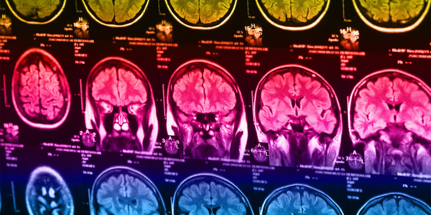
In their new study, Williams and her colleagues employed functional MRI (fMRI) to scan the brains of 801 participants diagnosed with depression or anxiety. These brain scans were performed both at rest and during tasks designed to probe cognitive and emotional functions, such as recognizing facial expressions and performing a Go–NoGo task, which tests cognitive control.
To refine their data, the researchers focused on six brain circuits known to be involved in depression: the default mode, salience, attention, negative affect, positive affect, and cognitive control circuits. They used the Stanford Et Cere Image Processing System to quantify brain activity and connectivity in these regions. The processing system ensured standardized measurements, which were expressed in standard deviation units relative to a healthy reference group.
The researchers identified six distinct biotypes of depression, each characterized by unique brain activity patterns.
“Our new brain scan biotype technology – The Stanford Et Cere Imaging Processing System – allows us to use fMRI to identify brain circuit dysfunctions in depression,” Williams told PsyPost. “By quantifying brain function at rest and during specific tasks we have shown that depression consists of six specific patterns of dysfunctions in six major brain circuits.”
The six biotypes were:
Biotype 1 (Overactivity in Cognitive Regions): Participants with this biotype exhibited overactivity in brain regions related to cognitive functions. This group showed the best response to the antidepressant venlafaxine (Effexor).
Biotype 2 (High Resting Activity in Three Brain Regions): This biotype was marked by higher levels of activity in three specific brain regions associated with depression and problem-solving when at rest. Participants in this group responded well to behavioral talk therapy.
Biotype 3 (Low Activity in Attention-Control Circuit): Characterized by lower activity at rest in the brain circuit that controls attention, this group was less likely to benefit from talk therapy compared to other biotypes.
Biotype 4 (Hyperconnectivity in Default Mode, Salience, and Attention Circuits): This biotype displayed intrinsic hyperconnectivity within these circuits, leading to slower emotional and attentional responses. Participants in this group responded better to a combined behavioral intervention.
Biotype 5 (Hypoconnectivity in Attention Circuit): Participants in this biotype showed significant lapses in concentration and impulsivity due to reduced intrinsic connectivity in the attention circuit.
Biotype 6 (Heightened Activity in Emotion Processing Circuits): This group exhibited increased activity in brain regions associated with processing both sad and positive emotions. Clinically, this was linked to severe anhedonia (loss of pleasure) and ruminative thinking.
“We were pleasantly surprised that the biotypes identified by our new fMRI technology were robust, meaning they were consistently observed even after multiple statistical validation tests,” Williams said. “We were also pleasantly surprised by the importance of task fMRI in identifying biotypes, in addition to resting state fMRI.”
To further refine their findings, the researchers conducted a randomized controlled trial with 250 participants. These participants were randomly assigned to one of three commonly used antidepressants (escitalopram, sertraline, or venlafaxine) or a behavioral talk therapy. This trial allowed the researchers to observe how different subtypes of depression responded to various treatments.
They found that these biotypes correlated with different clinical symptoms and treatment responses. For instance, the biotype characterized by overactivity in cognitive regions (Biotype 1) responded better to venlafaxine, while those with high resting activity in three brain regions (Biotype 2) benefited more from behavioral talk therapy. This differentiation suggests that specific brain activity patterns can predict which treatment will be most effective for individual patients.
“This discovery can be transformative,” Williams said. “It is a path for moving beyond diagnosis based only on observed symptoms, which is a one-size-fits all approach which doesn’t tell us which treatment will work best for each person. It opens the door for precision medicine in mental health.”
“We are also heartened by the potential of this approach to reduce stigma. We observe that when individuals can ‘see their brain,’ it gives a tangible way to understand what is happening for them, and why certain treatments are being considered. It removes the sense of blame and experience of ‘this is my fault, why can’t I control it.’”
“Although we had data on five different treatment conditions, it was not possible to consider all treatments available for depression that could be associated with specific biotypes,” Williams noted. “Our ongoing research is expanding to other treatments.”
For example, in another study published in Nature Mental Health, Williams’ team demonstrated the efficacy of transcranial magnetic stimulation (TMS) for individuals with the cognitive biotype of depression. The study involved 43 veterans, 26 of whom were identified as having the cognitive biotype through fMRI scans. After 30 daily TMS sessions targeting the cognitive control circuit, these veterans showed significant recovery in brain connectivity and improvement in cognitive control tests, with most benefits observed within the first five days of treatment.
While the findings represent a major step forward, the researchers acknowledge that more work is needed to refine these biotypes and validate them in larger, more diverse populations. Future studies could explore additional brain regions and functions, as well as the impact of different treatment modalities.
“Because the biotype approach is new to psychiatry and psychology, we don’t yet know how many biotypes will be important or necessary for encompassing all variations in depression and anxiety,” Williams explained. “Prior studies have reported two to four biotypes. With the addition of task fMRI data, we identified six biotypes. We did not quantify and analyze all known circuits of the brain and more biotypes could be identified by expanding coverage of additional circuits.”
“Our long-term goal is to move the field forward toward precision psychiatry,” Williams told PsyPost. “We are pursuing prospective trials in which we determine the most effective treatments for each biotype and apply them promptly to individual patients.”
Williams has received a five-year, $18.86 million grant from the National Institute for Health to develop a diagnosis and treatment tool for depressive disorders. The project will use brain imaging, computerized tests, and a novel smartphone app to create personalized cognitive biotypes for depression, aiming to transform diagnosis and treatment by providing tailored predictions and treatment guidance.
“We aim to scale up our research, testing more treatments across all six biotypes and using AI to refine and discover new biotypes,” Williams explained. “This expansion will connect brain imaging biotypes to clinical and digital measures, potentially allowing remote monitoring. At Stanford and other sites, we are already integrating imaging technology into clinical settings and hope to make it accessible to more clinicians and patients.”
Williams also highlighted the potential impact of the findings for young people.
“This precision approach based on brain scans has the potential to help address the global depression crisis,” she explained. “Around the world, 280 million people (about 8% of the population) will experience major depressive disorder. We commonly focus on the persistent low mood aspect of clinical depression. However, the other symptoms, anchored in different brain circuits, are bigger contributors to the disabling effect of the illness.”
“Unlike other chronic illnesses, it affects young people in the prime of their life. Without early effective treatment, it can have a chronic impact throughout their life, and dramatically increase years lost to disability. The vast majority of people cycle through many ‘failed’ treatment trials over many years. This process is associated with worsening disability and risk of suicide. Having a biotype test available early in the process has the potential to get many more people to the right treatment sooner, boosting the rate of recovery and life potential.”
The study, “Personalized brain circuit scores identify clinically distinct biotypes in depression and anxiety,” was authored by Leonardo Tozzi, Xue Zhang, Adam Pines, Alisa M. Olmsted, Emily S. Zhai, Esther T. Anene, Megan Chesnut, Bailey Holt-Gosselin, Sarah Chang, Patrick C. Stetz, Carolina A. Ramirez, Laura M. Hack, Mayuresh S. Korgaonkar, Max Wintermark, Ian H. Gotlib, Jun Ma, and Leanne M. Williams.


